Neuroimage. 2012 Nov 15;63(3):1561-70. doi: 10.1016/j.neuroimage.2012.07.060. Epub 2012 Aug 3.
Functional MRI-guided probabilistic tractography of cortico-cortical and cortico-subcortical language networks in children.
Broser PJ1, Groeschel S, Hauser TK, Lidzba K, Wilke M.
Abstract
In this study, we analyzed the structural connectivity of cortico-cortical and cortico-subcortical language networks in healthy children, using probabilistic tractography based on high angular resolution diffusion imaging. In addition to anatomically defining seed and target regions for tractography, we used fMRI to target inferior frontal and superior temporal cortical language areas on an individual basis. Further, connectivity between these cortical and subcortical (thalamus, caudate nucleus) language regions was assessed. Overall, data from 15 children (8f) aged 8-17 years (mean age 12.1 ±3 years) could be included. A slight but non-significant trend towards leftward lateralization was found in the arcuate fasciculus/superior longitudinal fasciculus (AF/SLF) using anatomically defined masks (p>.05, Wilcoxon rank test), while the functionally-guided tractography showed a significant lateralization to the left (p<.01). Connectivity of the thalamus with language regions was strong but not lateralized. Connectivity of the caudate nucleus with inferior-frontal language regions was also symmetrical, while connectivity with superior-temporal language regions was strongly lateralized to the left (p<.01). To conclude, we could show that tracking the arcuate fasciculus/superior longitudinal fasciculus is possible using both anatomically and functionally-defined seed and target regions. With the latter approach, we could confirm the presence of structurally-lateralized cortico-cortical language networks already in children, and finally, we could demonstrate a strongly asymmetrical connectivity of the caudate nucleus with superior temporal language regions. Further research is necessary in order to assess the usability of such an approach to assess language dominance in children unable to participate in an active fMRI study.
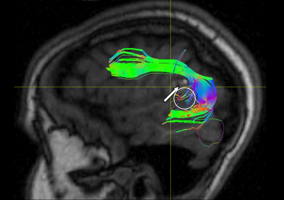
Figure 1: Example of a fMRI driven tractography of the F. Arcuatus in a patient with a small cortical lesion (white arrow) and abberant fibers (circle).
Figure 2: Functional guide fMRI of F. Arcuatus, Average over 15 subjects , F. Arcuatus on left (red) and right side (green).