
Aim To track the magnetic field generated by the propagating muscle action potential (MAP).
Method: In this prospective, proof of principle study, the magnetic activity of the intrinsic foot muscle after electric stimulation of the tibial nerve was measured using optically pumped magnetometers (OPMs). A classical biophysical electric dipole model of the propagating MAP was implemented to model the source of the data.
Results: The signal profile generated by the activity of the intrinsic foot muscles was measured by four OPM devices. Three devices were located above the same muscle to compare the direction and the strength of the magnetic signal while propagating along the muscle.
Interpretation: OPM devices allow for a new, non-invasive way to study MAP patterns. Since magnetic fields are less altered by the tissue surrounding the dipole source compared to electric activity, a precise analysis of the spatial characteristics and temporal dynamics of the MAP is possible. The classic electric dipole model explains major but not all aspects of the magnetic field. The field has longitudinal components generated by intrinsic structures of the muscle fibre. By understanding these magnetic components, new methods could be developed to analyse the muscular signal transduction pathway in greater detail.
The central nervous system exerts control over the activation of muscles via a dense network of nerve fibres targeting each individual muscle. There are numerous clinical situations were a detailed assessment of the nerve-innervation pattern is required for diagnosis and treatment. Especially deep muscles are hard to examine and are as yet only accessible by uncomfortable and painful needle EMG techniques. Just recently, a new and flexible method and device became available to measure the small magnetic fields generated by the contraction of the muscles: Optically pumped magnetometers (OPM). OPMs are small devices that measure the zero-field level crossing resonance of spin-polarized rubidium atoms. The resonance is dependent on the local magnetic field strength and therefore these devices are able to measure small magnetic fields in the range of a few hundred femto Tesla. In this study, we demonstrate as a proof of principle that OPMs can be used to measure the low magnetic fields generated by small hand muscles after electric stimulation of the ulnar or median nerve. We show that using this technique we are able to record differential innervation pattern of small palmar hand muscles and are capable of distinguishing between areas innervated by the median or ulnar nerve. We expect that the new approach will have an important impact on the diagnosis of nerve entrapment syndromes, spinal cord lesions and neuro muscular diseases.
Example:
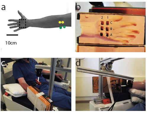
This figure shows the set up for the measument:
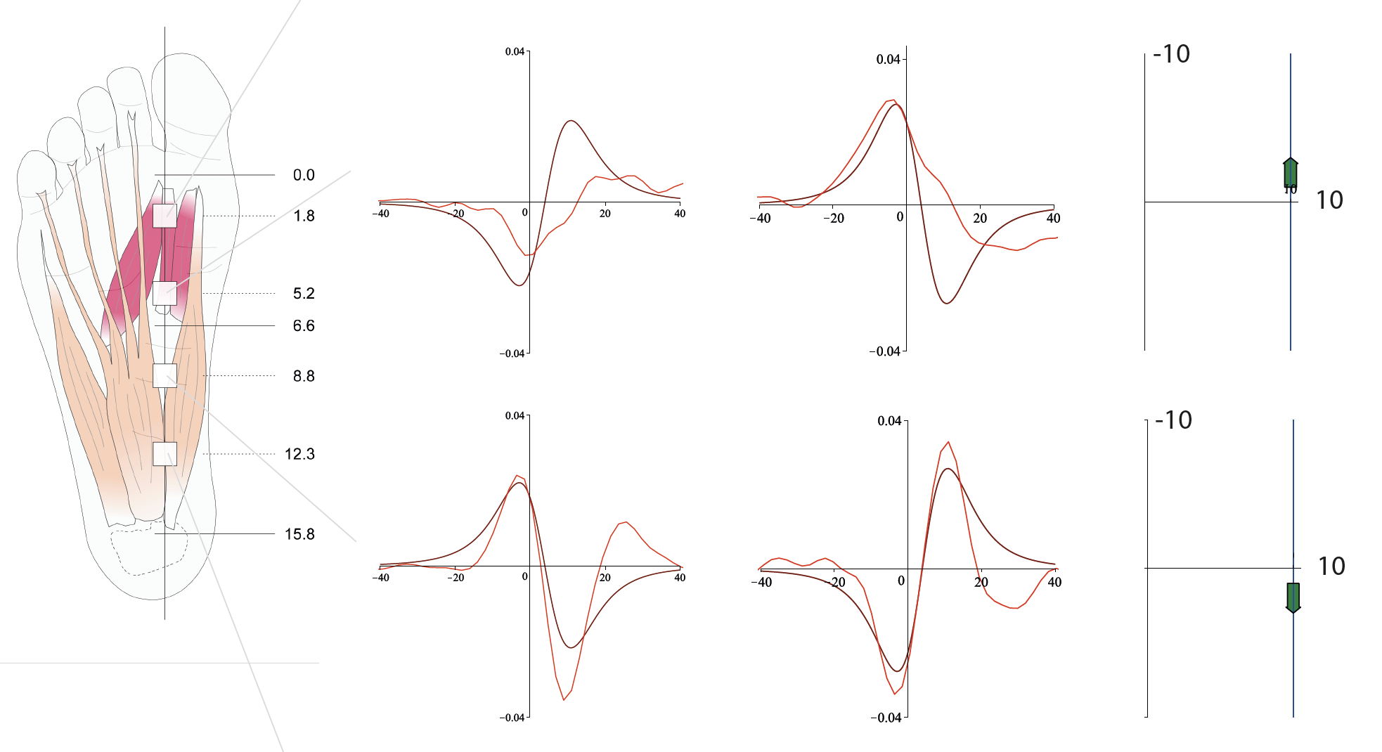
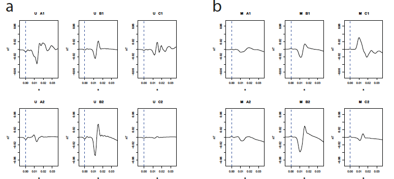
This figure shows the siganl profile recorded at the hand a) after median nerve stimulation, b) after ulnar nerve stimulation.
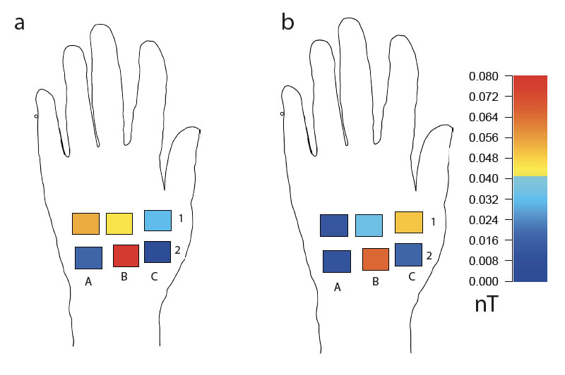
This figure shows the two distribution graphically visualized:
a) median nerve stimulation
b) ulnar nerve stimulation
see:
P. J. Broser, S. Knappe, D. S. Kajal, N. Noury, O. Alem, V. Shah, and
C. Braun. Optically Pumped Magnetometers for Magneto-Myography to
Study the Innervation of the Hand. IEEE Trans Neural Syst Rehabil Eng,
26(11):2226-2230, Nov 2018 (3.410)