Jenny, Lütschg Broser, 2020 ( Preprint )
During the first months of life the peripheral nerval system undergoes a dramatic maturation process. For instance, the nerve conduction speed (NCS) increases during the first two years from as low as 20m/s to almost the adult values of about 50m/s. How this increase in NCS is correlated to the structural maturation of the peripheral systems is hardly known. The objective of the study was therefore to analyse the maturation and growth of the cross-sectional area of the median nerve from birth to the first months of life using high resolution ultra-sound.
High-resolution ultrasonography of the median nerve was performed at three location in 43 infants and children without chronic diseases. The cross-sectional area (CSA) was measured and plotted against age and put in relation to the nerve conduction speed (NCS).
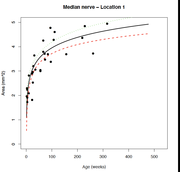
The high-resolution images demonstrated a dramatic maturation of the inner structure of the median nerve. At time of birth there is hardly any echogenic tissue present inside the nerve. At the age of two years one easily sees the fascicular structure of the nerve. Further the CSA increases significantly in a linear logarithmic relation to the age of the patient. The NCS correlates linearly with the CSA during the observed time period.
This study demonstrates the dramatic maturation of the peripheral nervous system during the first years of life. In addition, it provides normative data for the median nerve during the observed time period.
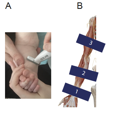
This image demonstrates how and at which locations the imaging was performed:
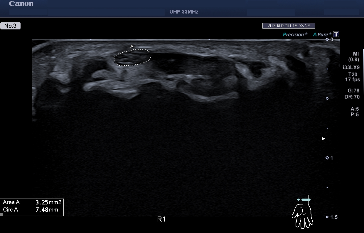
High resolution image of the median nerve at the level of the wrist (Location 1) of a child 1 month old.
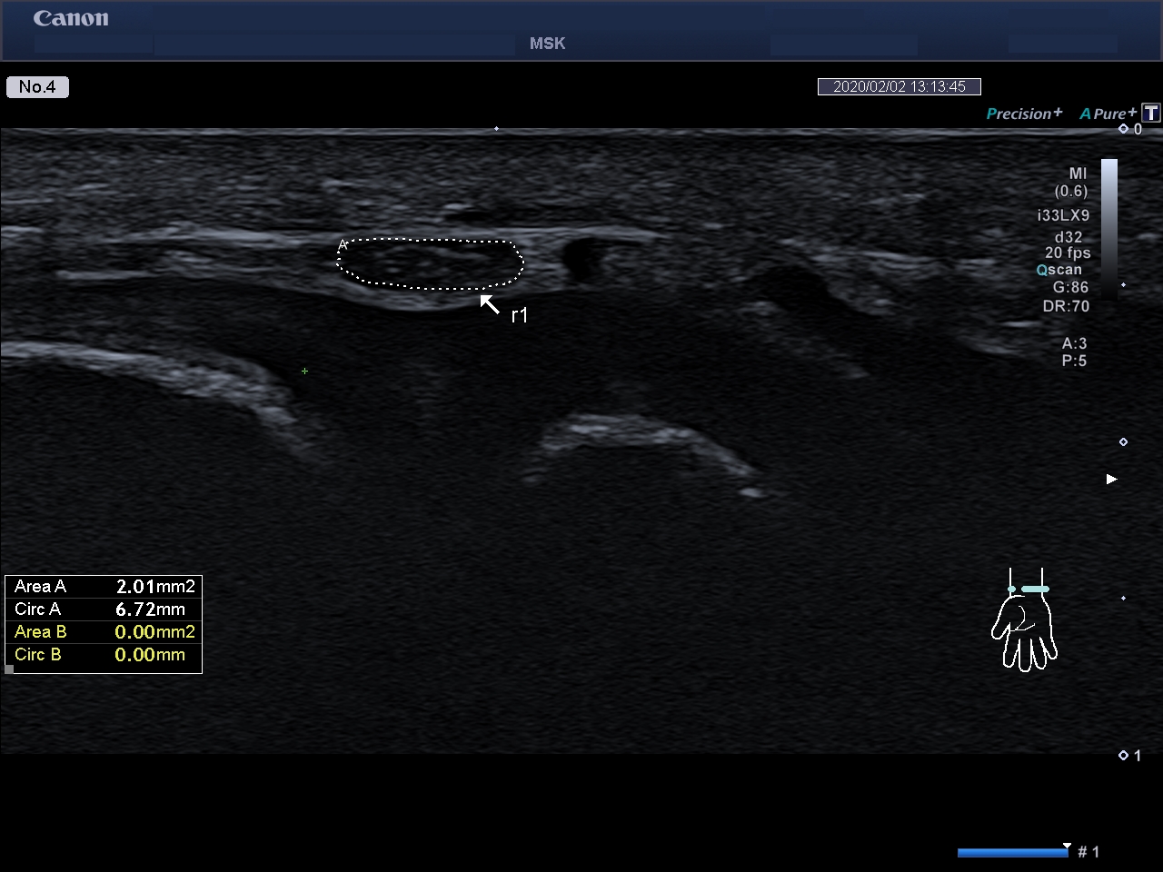
Second example of the n. medianus at the level of the writs (child 4 weeks)
The increase in the crossectional area was systematically measured. The increase follows a linear logarithmic curve: