AxoVis visualizes the Axonal Projection domain of Layer 2/3 somatosensory cortex interactively as described in
J Neurosci Methods. 2008 Mar 30;169(1):43-54. doi: 10.1016/j.jneumeth.2007.11.027. Epub 2007 Dec 8.
Automated axon length quantification for populations of labelled neurons.
Broser PJ1, Erdogan S, Grinevich V, Osten P, Sakmann B, Wallace DJ.
Abstract
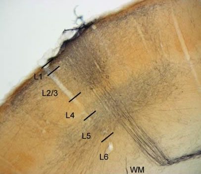
Virus-based methods for labelling populations of cortical neurons, when combined with cell-type specific recombinant promoters and techniques allowing temporal control of gene expression, provide neuroscience with new opportunities to examine the connectivity between brain regions and how this connectivity is modified by experience or disease. However, to take full advantage of these technical advances, it is necessary to develop new methods for quantification of the axonal projections revealed. Here we describe a method for quantitative analysis of axonal projection patterns emanating from populations of labelled cells, using transmitted light bright field microscopy. A single high resolution image of an area to be analysed is first acquired using mosaic extended focus image microscopy. This image is then analysed by specifically developed image processing algorithms that identify and track axon segments present. For quantitative analysis, measurement grids consisting of a user-defined number of individual elements are placed over an area of interest, with the computer-based method then returning the summed length of the axon segments in each element. Axon density plots can thus be generated. We present an example from rat brain showing, over a whole coronal section, axon densities emanating from a population of layer 2/3 somatosensory neurons.
The technique was used in the project:
Cereb Cortex. 2008 Jul;18(7):1588-603. Epub 2007 Nov 12.
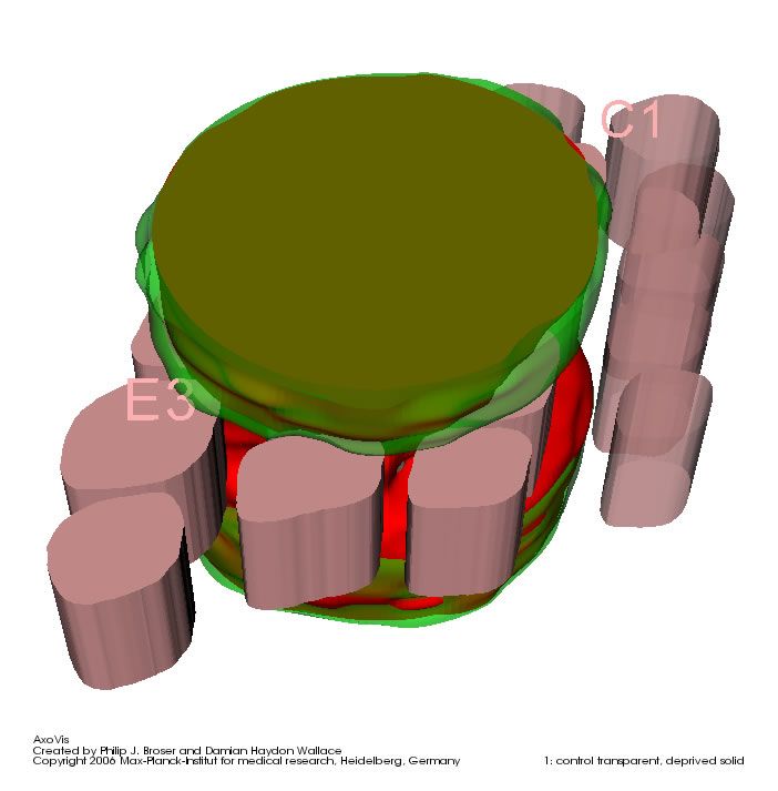
Critical period plasticity of axonal arbors of layer 2/3 pyramidal neurons in rat somatosensory cortex: layer-specific reduction of projections into deprived cortical columns.
Broser P1, Grinevich V, Osten P, Sakmann B, Wallace DJ.
Abstract
We examined the effect of whisker trimming during early postnatal development on the morphology of axonal arbors in rat somatosensory cortex. Axonal arbors from populations of layer 2/3 pyramidal neurons in the D2 column were labeled by lentivirus-mediated expression of green fluorescent protein. Axonal projection patterns were compared between untrimmed control animals and animals with all whiskers in A-, B-, and C-rows trimmed (D- and E-rows left intact) from postnatal days 7 to 15 (termed from here on DE-pairing). Control animals had approximately symmetrical horizontal projections toward C- and E-row columns in both supra- and infragranular layers. Following DE-pairing, the density of axons in supragranular layers projecting from the labeled neurons in the D2 column was higher in E- than in C-row columns. This asymmetry resulted primarily from a reduction in projection density toward the deprived C-row columns. In contrast, no change was observed in infragranular layers. The results indicate that DE-pairing during early postnatal development results in reduced axonal projection from nondeprived into deprived columns and that cortical neurons are capable of structural rearrangements at subsets of their axonal arbors.
Currently there is a version for Mac OSX and Windows available.