AxoQuant :
AxoQuant is a package of Unix Tools tools to filter and process fluorescence image data from neurons acquired with Two Photon or Con-focal Microscopy
in order to reconstruct the neuronal morphology and to measure ion channel distributions.
Front Neural Circuits. 2013 Apr 4;7:61. doi: 10.3389/fncir.2013.00061. eCollection 2013.
Three-dimensional tracking and analysis of ion channel signals across dendritic arbors.
Ginger M1, Broser P, Frick A.
Abstract
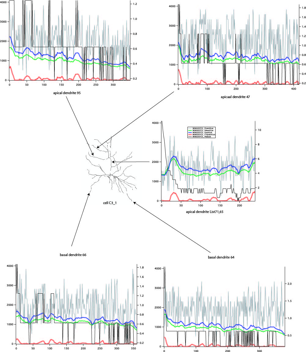
Most neuron types possess elaborate dendritic arbors that receive and integrate excitatory and inhibitory inputs from numerous other neurons to give rise to cell-type specific firing patterns. The computational properties of these dendrites are therefore crucial for neuronal information processing, and are strongly determined by the expression of many types of voltage-gated ion channels in their membrane. The dendritic distribution patterns of these ion channels are characteristic for each ion channel type, are dependent on the neuronal identity, and can be modified in a plastic or pathophysiological manner. We present a method that enables us to semi-automatically map and quantify in 3D the expression levels of specific ion channel types across the entire dendritic arbor. To achieve this, standard immunohistochemistry was combined with reconstruction and quantification procedures for the localization and relative distribution of ion channels with respect to dendritic morphology. This method can, in principle, be applied to any fluorescent signal, including fluorescently tagged membrane proteins, RNAs, or intracellular signaling molecules.
The tools are currently used for the project:
Three dimensional tracking of ion channels on epileptogenic cortical neurons in focal dysplasia in humans.
Background: About 20 Percent of children with focal epilepsy will be pharmacoresistant (1). Especially children with imaging abnormalities are most likely to be unresponsive to anti epileptic drug treatment. A significant number of abnormalities are due to focal cortical dysplasia. These focal epilepsies remain a major challenge to modern neuropediatrics and epileptic drug treatment. The mechanisms leading to a raised excitability in the dysplastic cortical tissue and its surrounding –the zone of irritation- is hardly understood. It is well known that the surface of neurons is packed with ion channels. These channels critically determine the firing pattern of the cell. The electro-chemical behavior of the cell is determined by the molecular structure of the channels, the quantities of ion channels and the exact distribution of the channels on the neuronal surface. It was recently shown in rodents that the distribution of A-Type Potassium channels is altered on pyramidal cells in proximity to an epileptogenic cortical lesion.A further class of channels which are possibly altered in cortical lesions are calcium channels. In the literature (2) a compelling evidence exists linking T-type calcium channels to seizure generation. However no rigorous study was conducted to investigate if the distribution of T-type channels is altered in structural epilepsy. This altered distribution of ion channels might contribute to the raised excitability of neurons in epileptogenic cortical areas.In our hospital we look after children with structural epilepsy. Some children with epilepsy are treated by surgical resection of the lesion. In our study we use the resected tissue after neuropathological assessment for further analysis.The aim of the study is to analyse the resected tissue with respect to the ion channel distribution and to compare the results with the results obtained from normal brain tissue. Children typically have a much shorter history of antiepileptic drug treatment before they undergo epileptic surgery. Therefore the effect of medication on the ion channel distribution is assumed to be limited. In recent years we have developed a method that allows the reconstruction of the geometry of the neurons and further the tracing of the distribution of specific ion channels along the dendritic arbor (3). We could show that the method is able to quantify the distribution of specific ion channels along the dendritic surface. We are specifically interested in layer V pyramidal neurons from temporal or frontal cortical areas which are most homogeneous in terms of ion channel distribution. In our study the distribution of A-type potassium and T-type calcium channels of pyramidal neurons from epileptogenic tissue is compared to unaltered neuronal tissue from children undergoing tumor resection (access tissue). We are now in a process of systematic clinical characterization of patients and consecutive tissue collection.
1) Epilepsia. 2013/05, Predicting pharmacoresistance in pediatric epilepsy. Wirrell EC.
2) Epilepsia. 2012/12, Dendritic ion channelopathy in acquired epilepsy. Poolos NP, Johnston D.
3) Front Neural Circuits. 2013/4 Three-dimensional tracking and analysis of ion channel signals across dendritic arbors. Ginger M, Broser P, Frick A.